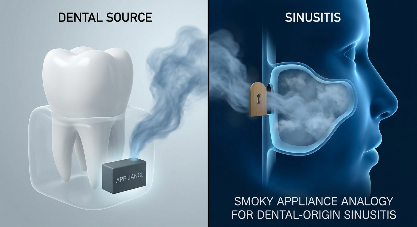

Sinus Mucocele Symptoms, Causes, and Treatment: Blocked Sinus (Mucocele) Explained
A sinus mucocele can sound like just another sinus problem, but it’s different from routine congestion. Understanding what it is—and when it deserves prompt attention—can help you know what to do next.
Quick Take: What a Sinus Mucocele Is (and Why It’s Different Than Congestion)
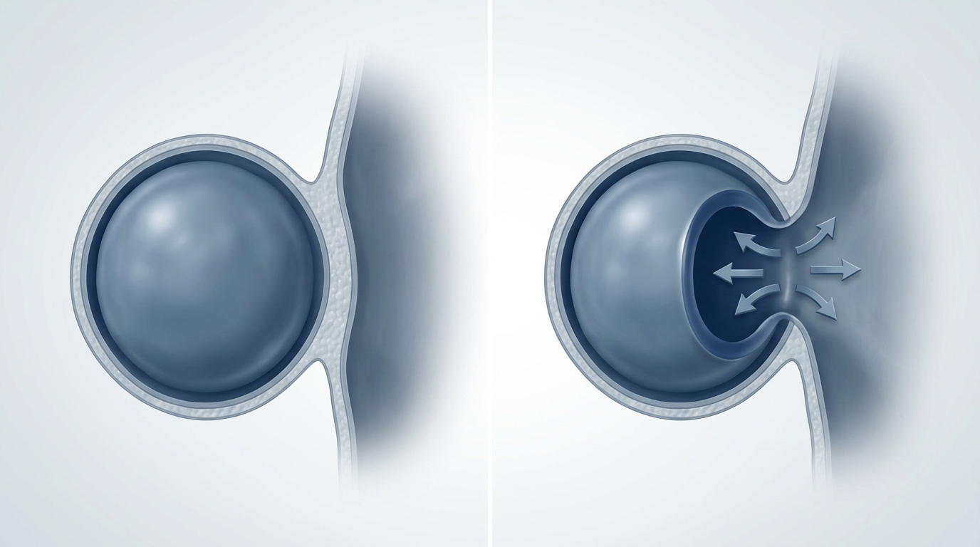
In plain language, a mucocele is a blocked sinus cavity that keeps filling with mucus, slowly expanding like a balloon. Because it can enlarge over time, it may thin or erode nearby bone and press on important structures such as the eye socket, nerves, or skull base. The reassuring news: most mucoceles are benign and slow-growing, but their location can make them medically significant.
A helpful analogy: typical congestion is like a clogged sink that still has somewhere to overflow. A mucocele is more like a closed container—fluid keeps accumulating, pressure builds, and the space can gradually stretch.
If sinus pressure lingers on one side or steadily worsens, it’s worth a closer look.

What Is a Sinus Mucocele?
Blocked sinus cyst explained
A mucocele is a mucus-filled, epithelium-lined expansion that forms when a sinus drainage pathway (ostium) gets obstructed. Think of each sinus as a room with a small drain. When the drain narrows or closes:
- Mucus can’t drain normally
- Mucus builds up
- Pressure increases
- The sinus may expand, remodeling or eroding the surrounding bony walls
This is why people sometimes describe a mucocele as a blocked sinus cyst—but it’s not simply a small pocket of fluid; it’s a sinus that can progressively enlarge.
One practical example: someone may have sinus pressure that doesn’t come and go like a cold. Instead, it lingers and slowly worsens, especially on one side, because the underlying issue is mechanical obstruction rather than a short-lived infection.
Common locations (and why location changes symptoms)
- Frontal and ethmoid sinuses: most common sites
- Sphenoid sinus: less common but taken seriously because it sits close to the optic nerve, carotid artery, and skull base
Location matters because it predicts what might get compressed. A frontal mucocele may feel like forehead pressure, while an ethmoid or sphenoid mucocele is more likely to involve the orbit (eye) or nearby nerves if it expands.
Mucocele vs. sinus retention cyst vs. nasal polyp (simple comparison)
- Mucocele: An obstructed sinus that expands with mucus; can remodel bone and affect the eye or nearby structures.
- Sinus retention cyst: A small mucus pocket in the sinus lining; often small and incidental; usually does not expand like a mucocele.
- Nasal polyp: A soft tissue growth related to inflammation; can cause blockage and congestion, often linked to chronic inflammation or allergies.
A mucocele is an obstructed sinus that expands over time, not just extra mucus.

Sinus Mucocele Causes and Risk Factors
The main cause: blocked drainage pathways
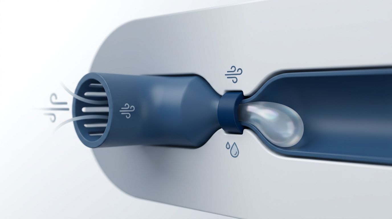
Most mucoceles start with obstruction of the sinus opening (ostium)—the natural drain that allows mucus to exit. The underlying issue is usually drainage failure, not too much mucus alone. Even normal mucus production becomes a problem if it can’t leave the sinus.
Common contributors
- Chronic inflammation, including chronic sinusitis (learn more: https://sleepandsinuscenters.com/chronic-sinusitis)
- Prior sinus surgery (scarring can reduce drainage)
- Trauma or facial fractures
- Tumors or masses that physically block drainage (uncommon, but important to rule out)
- Children: cystic fibrosis is a notable risk factor
A clinician may describe this as a plumbing issue: inflammation, scarring, or structural blockage narrows the outflow tract, and the sinus can’t ventilate or drain normally.
Who is more likely to develop one?
- Long-standing inflammatory sinus disease
- A history of sinus procedures or facial trauma
- Persistent symptoms that are one-sided or don’t match typical sinus infection patterns
For instance, a patient might say, I’ve had pressure behind my left eye for months, but antibiotics never really changed it. That kind of slow, one-sided pattern is one reason ENTs consider imaging.
Most mucoceles begin with a blocked drain; finding why it’s blocked helps guide treatment.
Sinus Mucocele Symptoms (By Location)
Symptoms can range from mild pressure to more urgent eye or vision issues—often depending on how far the mucocele extends beyond the sinus. A key theme: mucoceles often don’t behave like classic acute sinus infections. They may cause gradual, persistent symptoms rather than fever and sudden severe illness (unless infection complicates things).
Common symptoms (many overlap with sinus issues)
- Headache or facial pressure/pain
- Nasal congestion or a blocked feeling
- Recurring sinus symptoms that don’t behave like typical infections (for example, lingering or slowly worsening)
If symptoms are subtle, a mucocele may be found only after a CT scan done for chronic sinus pressure or for evaluation before or after sinus surgery.
Frontal sinus mucocele symptoms
- Forehead pressure/headache
- In more advanced cases, swelling or a contour change of the forehead or upper eye socket region
A concrete example: someone may notice that one eyebrow or upper eyelid area looks puffier over time, or that glasses feel different on one side due to gradual bony remodeling.
Ethmoid mucocele symptoms (near the eye socket)
- Eye pressure or discomfort, tearing, facial pain
- Proptosis (eye bulging) or double vision if there’s orbital involvement
When orbit-related symptoms appear, ENTs take them seriously because they can indicate extension beyond the sinus boundaries.
Sphenoid mucocele symptoms (deep sinus behind the nose)
- Deep headache, sometimes felt behind the eyes
- Vision changes due to proximity to the optic nerve
Because the sphenoid sinus is near critical neurovascular structures, imaging and treatment planning can be especially important in this location.
Steady, side-specific pressure or eye symptoms are clues it’s more than routine congestion.
Red Flags — When a Mucocele Might Be an Emergency
Some symptoms suggest orbital involvement or complications that deserve urgent evaluation. Seek urgent care for:
- New visual loss, blurred vision, or double vision
- Proptosis (eye bulging) or painful swelling around the eye
- Fever, severe or worsening headache, or neurologic symptoms (possible infection or extension)
New eye or vision symptoms should be treated as urgent until proven otherwise.
How a Sinus Mucocele Is Diagnosed
ENT evaluation (what to expect)
Diagnosis usually starts with an ENT visit that reviews:
- Symptom timeline (slow growth vs. sudden illness)
- Whether symptoms are one-sided
- History of sinus surgery, trauma, or chronic sinus disease
A nasal exam may include nasal endoscopy, where a thin camera is used to look inside the nasal passages and drainage areas in a detailed, well-lit view.
In clinic, ENTs are often listening for patterns like: This isn’t acting like a cold, or This keeps coming back in the same spot. Those clues help determine when imaging is the next best step.
Imaging is the key: CT first, MRI sometimes
Imaging is central because it shows whether a sinus is simply inflamed—or expanded and remodeling bone.
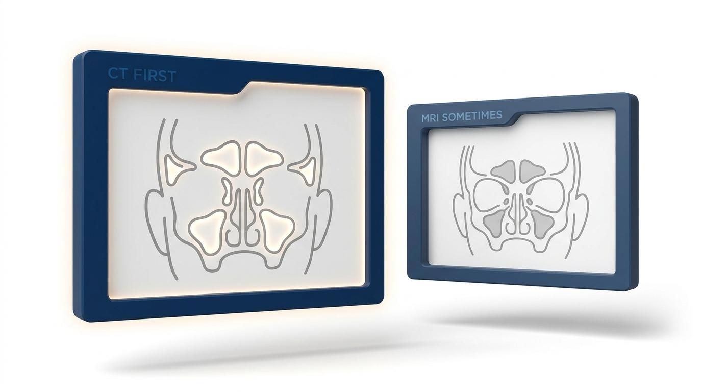
- CT scan = first-line test. CT typically shows complete sinus opacification along with expansion and thinning or defects of the bony walls, and it helps map anatomy for treatment planning. Related overview: https://sleepandsinuscenters.com/blog/sinus-ct-scan-what-it-shows-and-how-it-helps-diagnose-sinus-issues
- MRI = added when needed. MRI may be used to clarify orbital or intracranial extension, evaluate sphenoid disease, or help distinguish a mucocele from tumor-like alternatives.
Conditions doctors may need to rule out
Because some tumors and other inflammatory lesions can mimic similar findings, imaging—sometimes including MRI—helps ensure the diagnosis is accurate.
CT is the workhorse for diagnosis; MRI helps when critical structures are involved or the diagnosis is unclear.
Sinus Mucocele Treatment Options (What Actually Works)
Why true mucoceles usually need procedural treatment
With a true mucocele, the main issue is mechanical obstruction. Even if inflammation is present, mucus may continue accumulating if drainage isn’t restored. That’s why most cases eventually need a procedure rather than watch and wait. Timing and approach depend on size, location, symptoms, and surgical planning with an ENT.
It can help to think of it this way: sprays and rinses may calm surrounding inflammation, but they typically can’t reopen a completely blocked drainage pathway by themselves.
Standard of care: endoscopic drainage and marsupialization
The most common definitive approach is endoscopic treatment done through the nose (often without external incisions):
- The goal is to create a wide, permanent opening so mucus can drain freely
- This is often described as endoscopic marsupialization—opening the mucocele into the nasal cavity so it can continuously ventilate and drain
Outcomes are generally favorable, but follow-up is important. If you’d like a patient-friendly overview of what these procedures involve, see: https://sleepandsinuscenters.com/blog/endoscopic-sinus-surgery-what-patients-should-know
When more extensive surgery may be needed
- Challenging frontal sinus anatomy
- Large bony defects or significant extension
- Infection or recurrence after prior treatment
In selected situations, combined approaches (endoscopic plus external) may be considered.
Medications—what they can and can’t do
- Antibiotics: may be used when infection is present or suspected as a complication
- Steroid sprays or rinses: may reduce inflammation in the surrounding nasal or sinus lining, but they don’t remove a fixed blockage on their own
True mucoceles usually need a procedure to restore drainage; your ENT will tailor timing and approach to your situation.
Recovery, Follow-Up, and Risk of Recurrence
Why long-term follow-up is important
Recurrence can happen—especially if drainage remains impaired—and it may occur years later. That’s why long-term monitoring is often part of responsible care.
A practical way to frame follow-up: the goal is not only to fix today’s blockage, but to keep the drainage pathway open over time.
Post-treatment monitoring
Follow-up may include office visits, endoscopic checks, and sometimes repeat imaging depending on sinus location and extent. People are commonly advised to contact their surgical team if symptoms worsen after treatment—especially fever, increasing pain, or any eye or vision symptoms.
Fixing the blockage is step one; keeping the pathway open over time helps prevent recurrence.
Lifestyle Tips to Support Sinus Health (and Reduce Future Blockage Risk)
These steps don’t treat a mucocele by themselves, but they can support overall sinus health—particularly after treatment or in people prone to inflammation.
Support healthy drainage
- Saline irrigation as directed by your clinician (often used after sinus procedures)
- Maintaining humidity and hydration
- Avoiding irritants like smoke and strong fragrances that can aggravate nasal lining
Manage chronic sinus inflammation
- Address chronic rhinitis or sinusitis triggers (including allergies and irritants)
- Keep follow-ups if symptoms remain persistent—especially when they are one-sided
Healthy nasal lining and good drainage support long-term results after treatment.
FAQs About Sinus Mucoceles
Is a sinus mucocele cancer?
A sinus mucocele is typically benign. Imaging is used to confirm the diagnosis and rule out tumor-like alternatives.
Can a sinus mucocele go away on its own?
It rarely resolves without restoring drainage, because the opening stays blocked.
What does a mucocele feel like?
Many people report pressure or headache. Depending on location, it can also cause eye pressure, tearing, double vision, or other eye symptoms.
What’s the best scan for a suspected mucocele—CT or MRI?
CT is usually first-line, with MRI added when extension is unclear or when sphenoid disease or alternative diagnoses are concerns.
What happens if it’s not treated?
Without treatment, some mucoceles may keep enlarging and press on nearby structures (such as the eye socket), depending on location. An ENT can explain risks for your specific situation.
How likely is recurrence after surgery?
Recurrence is possible—even years later—so long-term follow-up is commonly recommended.
When to See an ENT (Next Steps)
Consider an ENT evaluation if you have sinus pressure or headaches that are persistent, one-sided, worsening, or associated with eye symptoms. For more guidance on timing and warning signs, see: https://sleepandsinuscenters.com/blog/when-should-i-see-an-ent
If you’d like help sorting out whether your symptoms fit a routine sinus issue or something that needs imaging, you can book an appointment here: https://www.sleepandsinuscenters.com/
If in doubt, get checked—especially for one-sided or eye-related symptoms.
Sources
1. Capra GG, Mullin DP. Paranasal Sinus Mucocele (2012). Otolaryngologic Clinics of North America. https://pmc.ncbi.nlm.nih.gov/articles/PMC3422585/
2. Bouatay R, et al. The Role of Imaging in the Management of Sinonasal Mucoceles (2019). https://pmc.ncbi.nlm.nih.gov/articles/PMC6850736/
3. Plantier D, et al. Mucocele: Clinical Characteristics and Outcomes in 46 Operated Patients (2018). https://pmc.ncbi.nlm.nih.gov/articles/PMC6331304/
4. Yale Medicine. Sinus Mucocele. https://www.yalemedicine.org/clinical-keywords/sinus-mucocele
5. Radiopaedia. Paranasal sinus mucocele. https://radiopaedia.org/articles/paranasal-sinus-mucocele-1?lang=us
This article is for educational purposes only and is not medical advice. Please consult a qualified healthcare provider for diagnosis and treatment.
If you have eye or vision changes, severe headache, fever, or rapidly worsening symptoms, seek urgent medical care.
Don’t let allergies slow you down. Schedule a comprehensive ENT and allergy evaluation at Sleep and Sinus Centers of Georgia. We’re here to find your triggers and guide you toward lasting relief.