Nasal Granulomas: Causes, Symptoms, and Treatment of Growths Inside Your Nose
Noticing a new growth inside your nose can be unsettling—especially if it crusts, bleeds, or makes one side feel blocked. Many people first notice something like a persistent “scab,” a sore spot that won’t heal, or a bump that seems to come and go. Sometimes it’s only obvious when you blow your nose and see recurring blood-tinged mucus or a crust that keeps reforming in the same exact place.
One important thing to know: “nasal granuloma” isn’t one single diagnosis. It’s a term often used to describe a pattern of chronic inflammation that can look like a lump, crusted area, or ulcer inside the nasal cavity. Because several different conditions can look similar—especially early on—a careful evaluation is often needed to sort out the cause [1,2].
In this article, you’ll learn:
- What “granuloma” means in plain English
- Common symptoms (including whole-body clues)
- Major causes—including autoimmune conditions and infections
- How ENT specialists evaluate these lesions
- Treatment options based on the underlying diagnosis—and when to seek prompt care
What Is a Nasal Granuloma?
Granuloma vs. “granulomatous inflammation” (plain-English explanation): A granuloma forms when the immune system tries to “wall off” something it perceives as a persistent problem—such as an infection, an irritant, a foreign material, or an immune-driven inflammatory process. This reaction is called granulomatous inflammation [2].
A helpful analogy: think of it like the body building a small “fence” around something it can’t easily clear. The “fence” is made of immune cells and inflammation, and it can change the shape and texture of the lining inside the nose over time [2].
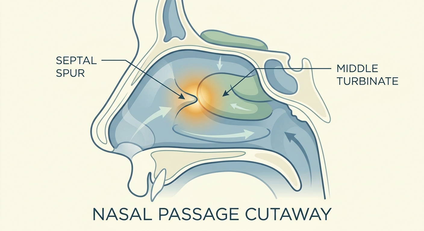
In the nose, granulomatous inflammation doesn’t always show up as a neat, round bump. It may appear as:
- A mass or growth
- A crusted area that repeatedly returns
- An ulcer or “raw spot”
- A polyp-like lesion
This variety is one reason nasal granulomas can be confusing at first glance—even for experienced clinicians without the benefit of endoscopy or tissue testing [2].
Nasal granuloma vs. nasal polyp vs. tumor (why appearance can mislead)
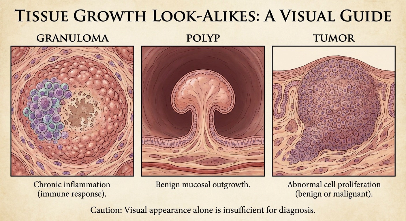
Different nasal conditions can look alike—especially without specialized examination. For example, a patient might say, “It looks like a polyp,” when what they’re actually seeing is a crusted ulcer or a fragile vascular growth that bleeds easily.
- Nasal polyps are typically soft, noncancerous inflammatory growths and often occur on both sides of the nose—but one-sided findings still deserve a full exam.
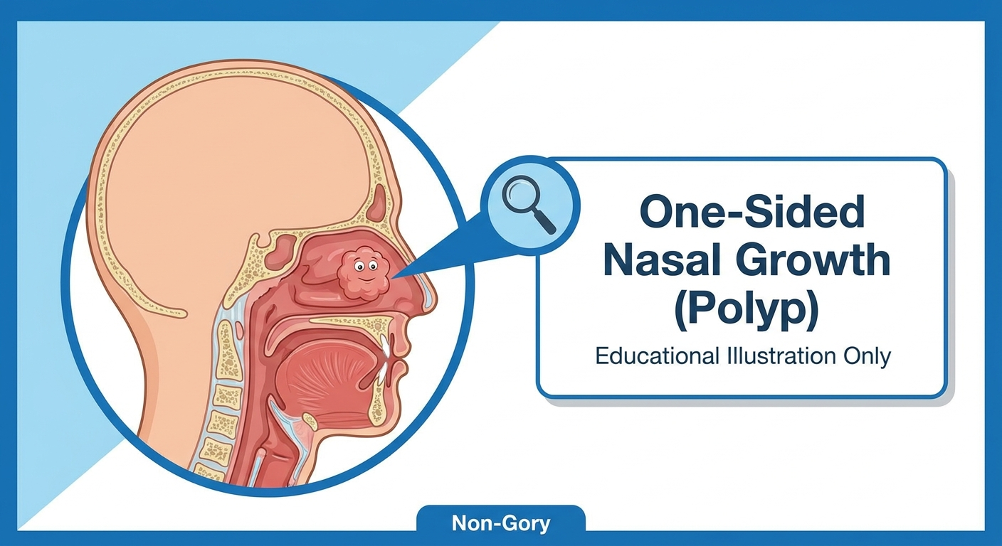
- Granulomatous lesions may involve crusting, ulceration, or a firm/irregular mass and may be one-sided.
- Tumors (benign or malignant) can also cause unilateral blockage and bleeding, among other causes—so persistent one-sided symptoms deserve evaluation [1].
It’s not possible to accurately diagnose nasal granulomas at home by looking—and it’s easy to mistake one type of nasal growth for another [1]. If you’re reviewing warning signs, this overview of early signs of nasal tumors can also be helpful in understanding why unilateral symptoms get attention: https://sleepandsinuscenters.com/blog/early-signs-of-nasal-tumors-key-symptoms-to-recognize

Common Symptoms of Nasal Granulomas (and Related Lesions)
Local (nose) symptoms
Symptoms can vary depending on the cause and the exact location, but commonly include:
- Persistent nasal blockage or congestion (often one-sided)
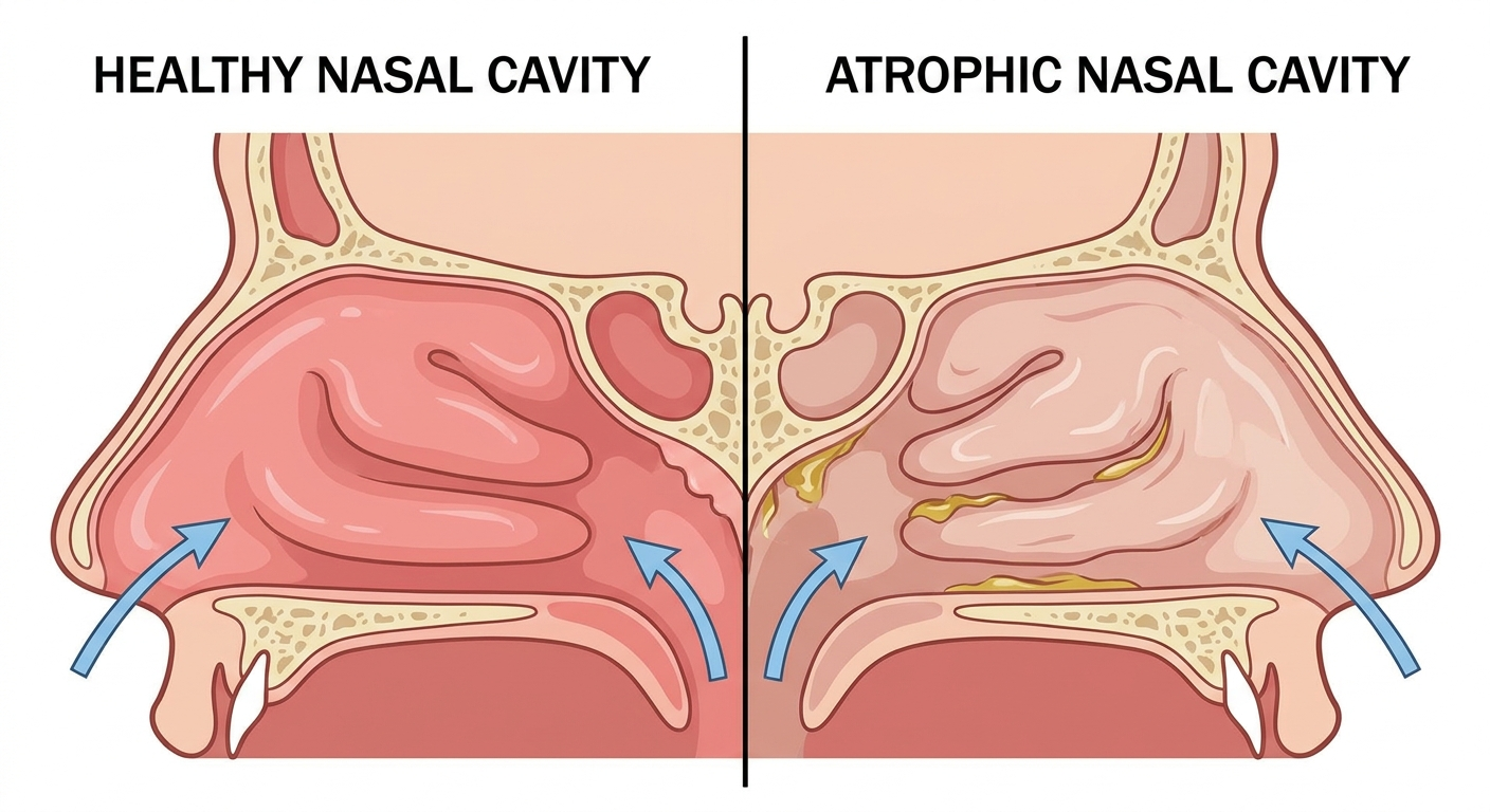
- Ongoing crusting or scabbing
- Nosebleeds or bleeding when wiping/blowing the nose
- A sore spot, ulcer, or “raw” area inside the nose
- Drainage (sometimes thicker or pus-like/purulent, depending on cause)
- Reduced sense of smell (in some cases)
A practical example: some people describe a “cycle” where the area crusts, they remove the crust to breathe better, it bleeds, and then it crusts again. That pattern doesn’t diagnose the cause—but it’s a useful detail to share with your ENT.
Symptoms that suggest a systemic condition (whole-body clues)
Sometimes, what looks like a local nasal issue is part of a broader inflammatory condition. Whole-body clues can include:
- Recurrent sinus infections and ongoing fatigue
- Cough, shortness of breath, or wheezing
- Joint pain, skin rashes, or eye irritation
- Fevers, night sweats, or unintentional weight loss
- Less commonly, coughing up blood or blood in the urine—details your clinician will want to know if present
These systemic symptoms matter because certain conditions associated with nasal granulomas—such as granulomatosis with polyangiitis (GPA) or sarcoidosis—can affect more than the nose and sinuses [3]. If you’ve noticed nose symptoms plus new symptoms elsewhere, mentioning the timing (what started first and what changed recently) can help guide the workup.
It’s helpful to view symptoms as clues that guide evaluation—not as a standalone diagnosis.
Causes of Growths Called “Nasal Granulomas”
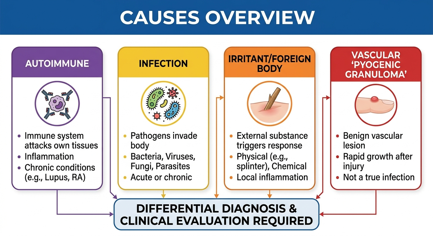
A “nasal granuloma” is a finding, not a final diagnosis. The underlying cause may range from autoimmune inflammation to infection to irritation or foreign material—and, less commonly, a neoplasm that needs to be ruled out [1,2].
Inflammatory/autoimmune causes (key categories)
Granulomatosis with polyangiitis (GPA)
GPA is an inflammatory disease that can affect small blood vessels and commonly involves the nose and sinuses. Symptoms may include:
- Crusting and chronic congestion
- Recurrent sinus infections
- Purulent drainage
- Nosebleeds [3]
When GPA-related inflammation is severe or untreated, it can damage cartilage and may contribute to problems such as septal perforation or changes in nasal structure [1,3]. Clinicians sometimes describe the nose as looking chronically irritated or friable, meaning it bleeds more easily with contact.
For a deeper look at sinus involvement, see: https://sleepandsinuscenters.com/blog/granulomatosis-with-polyangiitis-gpa-chronic-sinus-20260205051120
Sarcoidosis
Sarcoidosis is another inflammatory condition that can involve the nasal passages and sinuses. In the nose, it may mimic other chronic inflammatory or infectious problems—so symptoms alone rarely give a complete answer [1]. It is commonly considered in the “look-alike” group when granulomatous inflammation is seen.
Infections that can mimic autoimmune disease
Some infections can create granulomatous inflammation and look similar to autoimmune causes on exam:
- Fungal granulomas (certain fungi can trigger granulomatous inflammation)
- Tuberculosis (TB) involving the nose/sinuses (uncommon, but important to consider)
- Rhinosporidiosis, a chronic infection that can cause polyp-like lesions on nasal mucous membranes [1]
These infectious causes vary by region and exposure history. Because they can resemble inflammatory disease, confirming the cause often relies on targeted testing rather than appearance alone [1]. Two lesions can look similar in the office, but the correct treatment may be completely different—one might call for antimicrobial therapy, another for immune-directed medications [1].
Foreign body, irritants, trauma, and exposures
Inflammation can also develop when the nasal lining is repeatedly irritated or exposed to something it doesn’t tolerate well. Examples include:
- Retained foreign material (more common in children, but possible in adults)
- Ongoing irritation from smoke or occupational exposures
- Trauma or surgery-related irritation leading to chronic inflammation [1,2]
- Drug or toxic exposures that irritate or injure the nasal lining (considered based on history)
Sometimes the “cause” is less about a single event and more about ongoing irritation over time. An ENT might ask very specific questions here—work environment, hobbies, prior nasal procedures, and whether the area is repeatedly traumatized by picking or aggressive blowing—because that context can narrow the possibilities.
“Pyogenic granuloma” in the nose (important naming confusion)
A term that frequently causes confusion is pyogenic granuloma. Despite the name, it usually:
- Isn’t infectious
- Isn’t a true granuloma
In many cases, “pyogenic granuloma” refers to a benign vascular growth called a lobular capillary hemangioma, which can bleed easily. A common management approach is complete surgical excision—typically after confirmation of the diagnosis—to control bleeding and reduce recurrence risk, as advised by your surgeon [4].
If you’ve been told, “It’s a pyogenic granuloma,” it’s reasonable to ask a follow-up question: “Do you mean lobular capillary hemangioma?” That small clarification can help you understand why bleeding may be the main symptom and why removal is often recommended [4].
The same-looking nasal lesion can have very different causes—so diagnosis comes first, treatment second.
When to See an ENT (Red Flags You Shouldn’t Ignore)
Urgent/priority symptoms
It’s reasonable to seek prompt evaluation for symptoms such as:
- One-sided nasal blockage or a visible growth that persists
- Recurrent or heavy nosebleeds
- A crusted/ulcerated area that doesn’t heal
- Facial swelling, worsening pain, numbness, or any vision symptoms
- Unexplained weight loss, fevers, or night sweats
- Signs that could suggest septal perforation (for example, whistling with breathing, bleeding, crusting)
A simple rule of thumb: if a nasal problem is persistent, unilateral, and bleeding or ulcerating, it’s worth getting checked rather than watching it for “just a little longer” [1].
Why early evaluation matters
Different causes of nasal granulomas can require very different treatments—such as antibiotics, antifungals, immune-directed medications, or procedures. Getting the diagnosis right early helps guide the safest next step and can help prevent ongoing tissue injury in conditions that are potentially destructive [1,3].
When symptoms persist, prompt evaluation helps protect nasal structure and directs you to the right treatment sooner.

How Nasal Granulomas Are Diagnosed
ENT exam + nasal endoscopy
A typical evaluation starts with an ENT exam. Often, the next step is nasal endoscopy, which uses a small camera to view areas deeper in the nasal cavity that can’t be seen well otherwise.
During endoscopy, the clinician may assess:
- Where the lesion is located
- Whether it’s ulcerated or crusted
- Whether it bleeds easily
- Whether disease appears unilateral or bilateral
- Whether there are features that warrant further workup
If you want to know what to expect, this guide walks through the process in patient-friendly terms: https://sleepandsinuscenters.com/blog/what-is-nasal-endoscopy----and-is-it-painful
Imaging (when needed)
In some situations, imaging may be recommended—often a CT scan of the sinuses—to understand the extent of inflammation, whether bone/cartilage changes are present, and whether a deeper mass is suspected. Imaging doesn’t replace tissue diagnosis when that’s needed, but it can add important context—especially when symptoms suggest deeper sinus involvement.
Biopsy (often the key step)
Because many different conditions can look similar, a biopsy is often the most direct way to identify the cause and guide treatment. Tissue may be evaluated for specific inflammatory patterns, infection, or other abnormalities [1,5].
It can help to think of biopsy as moving from “what it looks like” to “what it is.” That distinction matters when the same visible finding (crusting, bleeding, a unilateral mass) could represent very different underlying diseases [1,2].
For a step-by-step overview, see: https://sleepandsinuscenters.com/blog/what-happens-during-a-sinus-biopsy
Lab tests and specialist referrals (based on suspected cause)
Depending on findings, additional evaluation may include:
- Bloodwork when conditions like GPA are suspected (often coordinated with rheumatology)
- Cultures and special stains from biopsy tissue when infection is a concern
Not everyone needs every test. Typically, the exam, endoscopy findings, and biopsy results determine what’s appropriate next.
Endoscopy, imaging, and—most importantly—biopsy help move from guesswork to a confident diagnosis.
Treatment Options (Based on the Underlying Cause)
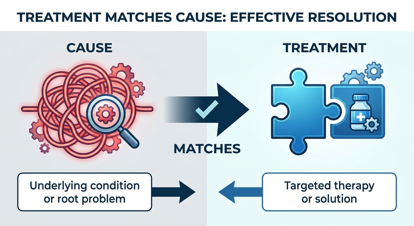
A helpful rule is: treat the cause—not just the appearance of the growth [1]. Managing nasal granulomas effectively depends on the diagnosis driving the inflammation.
If the cause is GPA (vasculitis)
Treatment for GPA typically involves systemic medications to control inflammation and prevent tissue damage, coordinated with appropriate specialists. Local care may also help with crusting or bleeding, but controlling the underlying inflammation is central [3].
If the cause is sarcoidosis
Sarcoidosis treatment depends on severity and which areas are involved. Some cases are monitored, while others use anti-inflammatory therapies [1]. The right plan is individualized—and often depends on symptoms, extent of involvement, and overall health.
If the cause is fungal disease, TB, or rhinosporidiosis
When infection is confirmed, management may include:
- Antifungal or antimycobacterial medications when indicated
- Procedural treatment in selected cases (for example, if a lesion is obstructive or prone to recurrence) [1]
Because infections can mimic inflammatory disease, it’s generally safest to focus on diagnosis first rather than trying to self-treat [1].
If the lesion is a “pyogenic granuloma” (lobular capillary hemangioma)
When the diagnosis is lobular capillary hemangioma, complete excision is commonly recommended to control bleeding and reduce recurrence risk; your ENT or surgeon will confirm whether that approach fits your case [4].
Different causes require different treatments—getting the diagnosis right is the fastest path to the right care.
Symptom Relief While Awaiting Diagnosis (supportive measures, not a treatment plan)
While evaluation is in progress, these are general comfort measures some clinicians suggest—not a cure or a substitute for professional evaluation:
- Saline spray or rinse for crusting (if tolerated). If using a rinse device, use only distilled, sterile, or boiled-then-cooled water, and follow CDC or device instructions.
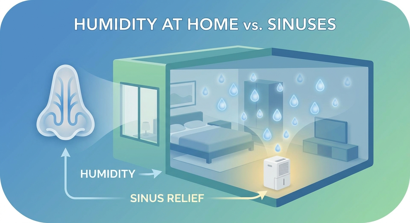
- Humidification and gentle nasal care (avoid picking).
- Avoiding smoke and harsh irritants.
If you’re bleeding easily, be especially gentle. Even helpful habits—like frequent swabbing or scraping crusts—can keep the cycle going.
Supportive care can improve comfort, but it won’t replace diagnosis-driven treatment.
Lifestyle Tips to Reduce Irritation and Support Healing (Adjuncts, Not a Cure)
Nose-friendly daily habits
- Use a humidifier if indoor air is dry and stay hydrated.
- Consider gentle saline rinses if your clinician agrees, following proper water-safety steps (distilled, sterile, or boiled-then-cooled water).
- Use only moisturizers or topical products recommended by your ENT team—some substances are not safe to place deep in the nose.
What to avoid
- Repeated trauma (picking, aggressive blowing).
- Overusing decongestant sprays (can lead to rebound congestion).
- Placing unknown “natural antimicrobials” in the nose, which can irritate or burn nasal tissue.
Small daily choices can reduce irritation while you and your clinician address the underlying cause.
FAQs About Nasal Granulomas
Are nasal granulomas cancer?
Not necessarily. Many are inflammatory or infectious. However, some cancers can present with similar symptoms (especially persistent, one-sided bleeding or blockage), which is why ENT evaluation and sometimes biopsy are important [1].
What’s the difference between a nasal granuloma and a pyogenic granuloma?
A “pyogenic granuloma” usually refers to a benign vascular growth (lobular capillary hemangioma). It’s typically not infectious and not a true granuloma, despite the name [4].
Why do doctors recommend a biopsy?
Because multiple diseases can produce similar-looking nasal lesions. A biopsy can clarify the cause and guide treatment choices [1,5].
Can a nasal granuloma go away on its own?
It depends on what’s driving it. Irritation-related inflammation may improve if triggers resolve, while autoimmune conditions or chronic infections often require targeted treatment.
What are signs something more serious is going on (like GPA)?
Ongoing crusting or bleeding, recurrent sinus infections, worsening nasal damage, and symptoms outside the nose (lungs, kidneys, joints) can raise concern for systemic disease and prompt further evaluation [3].
Key Takeaways
- “Nasal granulomas” describe a pattern of inflammation, not one single diagnosis [1,2].
- Important causes include GPA and sarcoidosis, plus infections like fungal disease, TB, or rhinosporidiosis (rarity varies by region/exposure) [1,3].
- Because several look-alike conditions exist, ENT evaluation with endoscopy and often biopsy is a common pathway to the correct diagnosis and treatment plan [1,5].
Next Step: Schedule an ENT Evaluation
If you’re dealing with persistent nosebleeds, crusting that won’t heal, or a one-sided nasal growth, an ENT evaluation can help clarify what’s going on and what options make sense.
To request a visit at Sleep and Sinus Centers of Georgia, you can book an appointment here: https://sleepandsinuscenters.com/appointments
This article is for educational purposes only and is not medical advice. Please consult a qualified healthcare provider for diagnosis and treatment. If you have concerning symptoms—especially heavy bleeding, vision changes, severe facial pain/swelling, or rapidly worsening symptoms—seek urgent medical care.
References
1. Fuchs FD, et al. Granulomatous disorders of the nose and paranasal sinuses. 2009. https://pubmed.ncbi.nlm.nih.gov/19225301/
2. Shah KK, et al. Histopathologic review of granulomatous inflammation. 2017. https://pmc.ncbi.nlm.nih.gov/articles/PMC6850266/
3. Mayo Clinic. Granulomatosis with polyangiitis—Symptoms and causes. 2025. https://www.mayoclinic.org/diseases-conditions/granulomatosis-with-polyangiitis/symptoms-causes/syc-20351088
4. Pathology Outlines. Lobular capillary hemangioma (pyogenic granuloma). 2024. https://www.pathologyoutlines.com/topic/skintumornonmelanocyticpyogenicgranuloma.html
5. UCSF Health. Nasal mucosal biopsy. 2025. https://www.ucsfhealth.org/care/medical-tests/nasal-mucosal-biopsy
Don’t let allergies slow you down. Schedule a comprehensive ENT and allergy evaluation at Sleep and Sinus Centers of Georgia. We’re here to find your triggers and guide you toward lasting relief.